In recent years, significant advancements in cardiac imaging and testing have revolutionized the diagnosis and management of cardiovascular diseases. These cutting-edge technologies and techniques provide healthcare professionals with a clearer and more detailed view of the heart, enabling early detection, precise diagnosis, and personalized treatment plans. This article explores the latest advances in cardiac imaging and testing, highlighting their importance in improving patient outcomes and enhancing the understanding of heart conditions. By staying informed about these breakthroughs, healthcare providers can offer the best possible care to patients with cardiovascular issues.
Cardiac Magnetic Resonance Imaging (MRI)
Cardiac MRI is a powerful imaging tool that provides detailed images of the heart’s structure and function. It utilizes a focused magnetic field and radio waves to create clear and high-resolution images of the heart, including the chambers, valves, blood flow, and surrounding tissues. Cardiac MRI is a non-invasive test and does not involve radiation, making it a safe option for diagnosing various heart conditions, such as heart muscle damage, cardiomyopathies, and congenital heart defects.
Coronary Computed Tomography Angiography (CCTA)
CCTA is a specialized imaging technique that allows for a comprehensive assessment of the coronary arteries, which supply blood to the heart muscle. This non-invasive test uses a computed tomography (CT) scanner and a contrast dye to visualize the coronary arteries, detecting blockages or narrowing that may cause chest pain (angina) or lead to a heart attack. CCTA is particularly valuable for patients with suspected coronary artery disease and provides detailed information that can guide treatment decisions.
Echocardiography
Traditional echocardiography uses sound waves to create two-dimensional images of the heart. Recent advancements in technology have allowed for the ability to gather further data that could help evaluate the heart’s anatomy and function. For example, 3D echocardiography provides a more quantitative and comprehensive evaluation of the heart’s pumping abilities. In addition, strain imaging can evaluate individual heart region’s pumping and stretching ability. Furthermore, if necessary, agitated saline can be injected to the blood stream to evaluate for a hole in the heart, or specialized lipid microbubbles can be administered to allow for a higher level image acquisition of the heart’s structures and function. These tools provide cardiologists with more specialized data of a person’s heart to better inform and treat patients.
Nuclear Cardiology
Nuclear cardiology combines nuclear medicine with imaging techniques to assess heart function and blood flow. One common nuclear cardiology test is the Myocardial Perfusion Imaging (MPI), which uses a small amount of radioactive tracer to show how well blood flows through the heart muscle during stress and rest. This test can help diagnose coronary artery disease and evaluate the extent of heart damage after a heart attack. Another more recent advancement in nuclear cardiology now allows us to conduct a nuclear test, if indicated, to evaluate for extra specialized protein in the heart.
Wearable Cardiovascular Devices
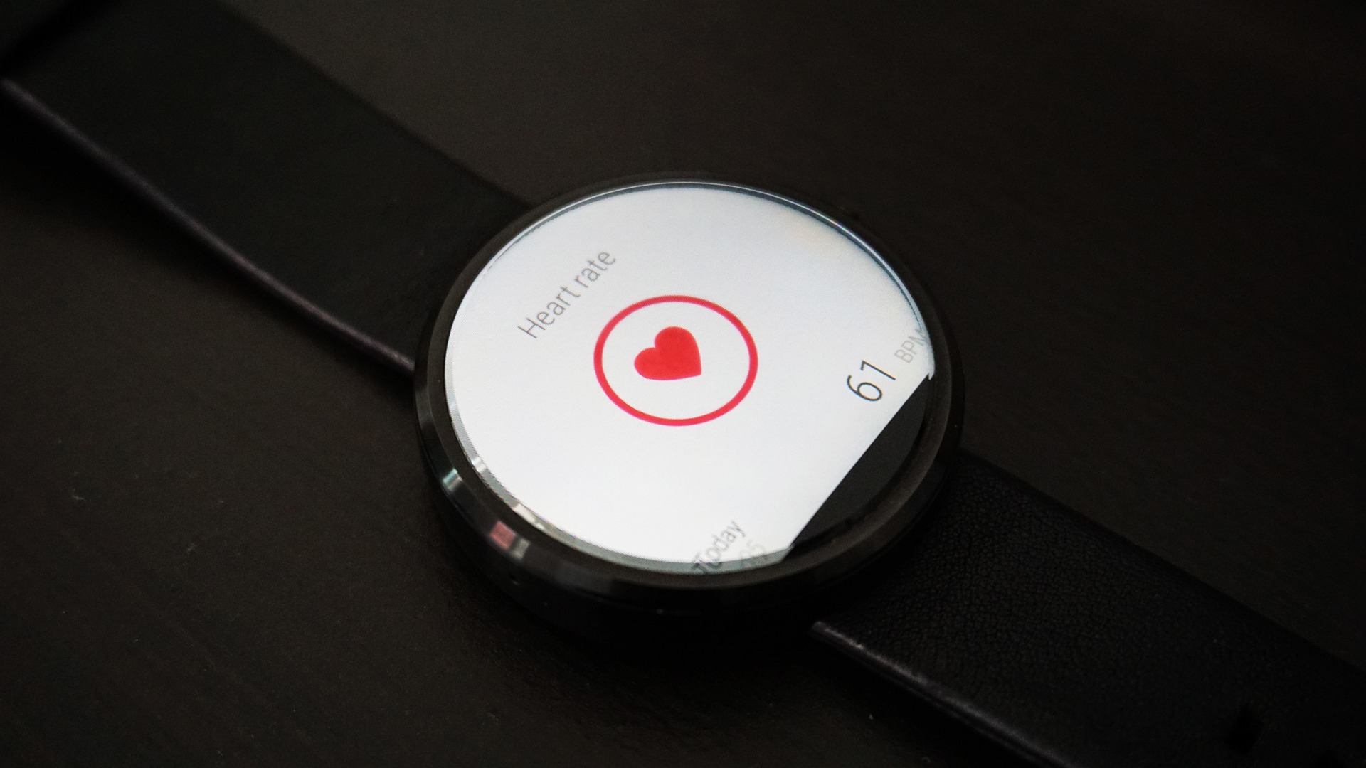
Advancements in wearable technology have introduced innovative cardiovascular devices that provide real-time monitoring of heart health. These wearable devices, such as smartwatches and fitness trackers, can measure heart rate, rhythm, and activity levels continuously. There are multiple FDA approved devices which could be helpful, in the right circumstances, in evaluating and treating patients with multiple subtypes of heart conditions. We advise working with your cardiologist to determine which device is best for you.
The latest advances in cardiac imaging and testing have ushered in a new era of cardiovascular care, providing healthcare professionals with sophisticated tools to accurately diagnose, monitor, and treat heart conditions. Cardiac MRI, CCTA, Advanced echocardiography, nuclear cardiology, and wearable devices have significantly improved the precision and efficiency of cardiac evaluations. As medical technology continues to evolve, these advancements have already begun to play a pivotal role in shaping the future of cardiovascular medicine and ultimately improving patient outcomes.
Sources:
- American Heart Association (AHA). (2021). Cardiac MRI. https://www.heart.org/en/health-topics/cardiac-mri
- National Heart, Lung, and Blood Institute (NHLBI). (2021). Cardiac Computed Tomography (CT) Scan. https://www.nhlbi.nih.gov/health-topics/cardiac-computed-tomography-ct-scan
- American College of Cardiology (ACC). (2021). 3D Echocardiography. https://www.acc.org/tools-and-practice-support/clinical-toolkits/3d-echocardiography
- Society of Nuclear Medicine and Molecular Imaging (SNMMI). (2021). Nuclear Cardiology. https://www.snmmi.org/clinical/nuclear-cardiology
- Rajpurkar, P. et al. (2018). Deep Learning for Cardiac Image Segmentation: A Review. Journal of Cardiovascular Magnetic Resonance, 20(1), 94. doi:10.1186/s12968-018-0519-6.
- Elgendi, M. (2019). Wearable Cardiac Devices. Sensors, 19(18), 4008. doi:10.3390/s19184008.


Leave A Comment